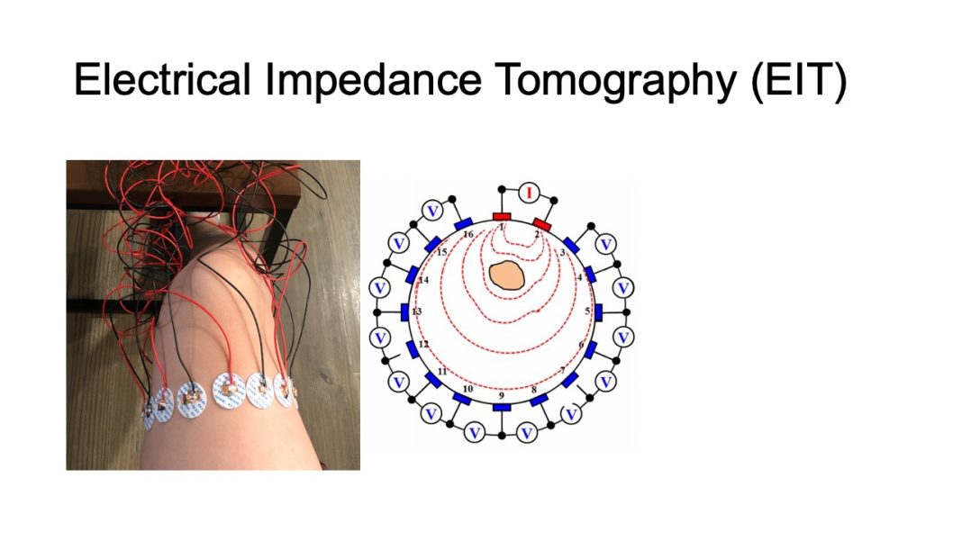
In the realm of medical imaging, a groundbreaking innovation is transforming our understanding of the brain and its functions. Electrical Impedance Tomography (EIT), traditionally known for applications in lung imaging and industrial processes, is now making waves as a pioneering method for imaging brain function. This article explores the recent medical breakthroughs in utilizing Electrical Impedance Tomography for imaging the brain and its potential implications for neuroscience and healthcare.
1. Traditional Brain Imaging Techniques:
Historically, imaging the brain has been dominated by techniques like Magnetic Resonance Imaging (MRI) and Functional Magnetic Resonance Imaging (fMRI). While these methods provide valuable structural and functional information, they often come with limitations such as cost, time consumption, and, in some cases, restricted mobility during scanning.
2. The Rise of Electrical Impedance Tomography:
2.1. Real-Time Imaging:
One of the standout features of EIT is its ability to offer real-time imaging. Unlike fMRI, which captures snapshots of brain activity over seconds, EIT provides continuous monitoring, enabling researchers to observe dynamic changes in brain function with unprecedented temporal resolution.
2.2. Non-Invasive Nature:
EIT’s non-invasive nature is a game-changer in brain imaging. With no exposure to ionizing radiation or strong magnetic fields, EIT allows for repeated imaging sessions without potential harm, making it particularly suitable for pediatric and long-term monitoring applications.
3. Applications in Neurological Research:
3.1. Seizure Monitoring:
EIT has shown promise in monitoring and mapping brain activity during epileptic seizures. The real-time capabilities of EIT enable clinicians to visualize the spread of abnormal electrical activity in the brain and tailor treatment approaches accordingly.
3.2. Neurocritical Care:
In neurocritical care settings, where continuous monitoring is crucial, EIT provides a valuable tool for assessing cerebral blood flow, detecting ischemic events, and guiding interventions to optimize brain function in real-time.
4. Advancements in Brain-Computer Interfaces:
4.1. Cognitive Function Mapping:
EIT holds potential for mapping cognitive functions in the brain. Researchers are exploring its use in understanding memory, attention, and language processing, paving the way for applications in brain-computer interfaces and neurorehabilitation.
4.2. Neuromodulation Guidance:
In neuromodulation therapies for conditions like Parkinson’s disease or depression, EIT could serve as a guiding tool for precise electrode placement and real-time assessment of treatment efficacy.
5. Future Perspectives and Challenges:
5.1. Integration with Other Modalities:
Future research aims to integrate EIT with other imaging modalities, such as MRI, to combine the strengths of each technique for a more comprehensive understanding of brain structure and function.
5.2. Addressing Spatial Resolution:
Challenges in spatial resolution persist, and ongoing research focuses on refining algorithms and electrode configurations to enhance the detail and accuracy of EIT brain images.
6. Potential Impact on Healthcare:
The ability to monitor brain function in real-time has transformative implications for patient care. EIT’s application in neurological research holds promise not only for diagnostics but also for guiding treatments, optimizing interventions, and personalizing therapeutic approaches.
In conclusion, the integration of Electrical Impedance Tomography into brain imaging represents a significant leap forward in understanding and treating neurological conditions. The real-time capabilities, non-invasive nature, and potential for continuous monitoring position EIT as a revolutionary tool in neuroscience, opening new avenues for research, diagnosis, and personalized treatment strategies in the realm of brain health.


